A Coronary Angiography is a procedure that uses X-ray imaging to see your heart’s blood vessels. Coronary angiograms are part of a general group of procedures known as heart catheterization. Heart catheterization procedures can both diagnose and treat heart and blood vessel conditions. A coronary angiogram, which can help diagnose heart conditions, is the most common type of heart catheterization procedure.
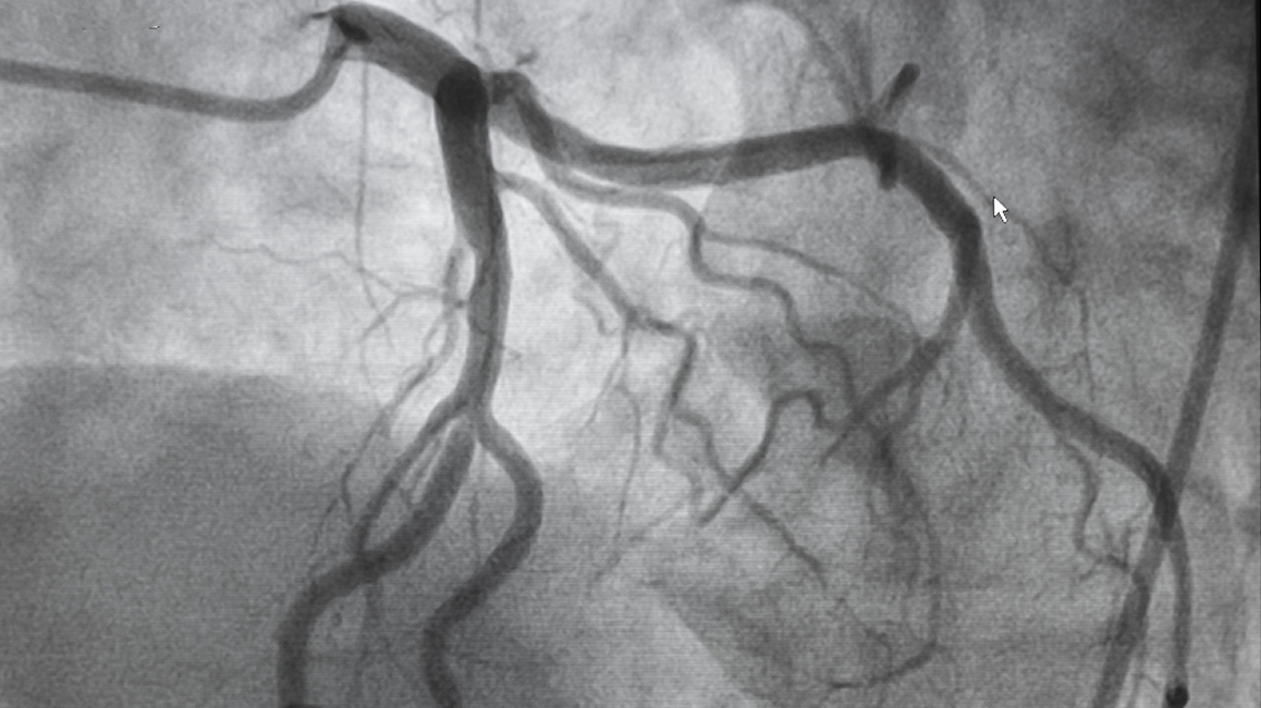
During a coronary angiogram, a type of dye that’s visible by an X-ray machine is injected into the blood vessels of your heart. The X-ray machine rapidly takes a series of images (angiograms), offering a detailed look at the inside of your blood vessels. If necessary, your doctor can perform procedures such as an angioplasty during your coronary angiogram.
A coronary angiogram is a special X-ray test. It’s done to find out if your coronary arteries are blocked or narrowed, where and by how much. An angiogram can help your doctor see if you need treatment such as Coronary Angioplasty or stent, coronary artery bypass surgery (CABG) or medical therapy.
Why do I need Coronary Angiography?
Coronary angiography can be used to help diagnose heart conditions, help plan future treatments and carry out certain procedures. For example, it may be used:
– after a heart attack – where the heart’s blood supply is blocked
– to help diagnose angina – where the pain in the chest is caused by the restricted blood supply to the heart
– to plan interventional or surgical procedures – such as a coronary angioplasty, where narrowed or blocked blood vessels are widened
Coronary angiography is also considered to be the best method of diagnosing coronary heart disease (where a build-up of fatty substances in the coronary arteries affects the heart’s blood supply).
What happens during Coronary Angiography?
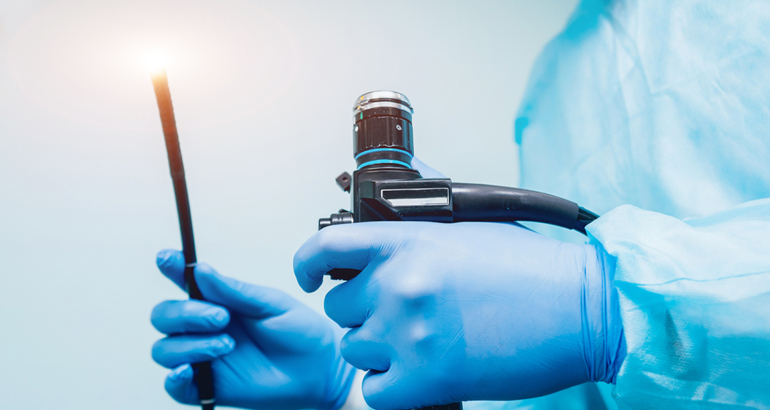
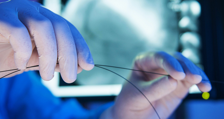
During the procedure, a long, thin and flexible tube called a catheter is inserted into a blood vessel from your groin or arm. Using X-ray images as a guide, the tip of the catheter is passed up to the heart and coronary arteries. A special type of dye called a contrast medium is injected into the catheter and X-ray images (angiograms) are taken. The contrast medium is visible on the angiograms, showing the blood vessels that the fluid travels through. This clearly highlights any blood vessels that are narrowed or blocked. The procedure is usually carried out under local anesthetic, so you will be awake while the procedure is carried out, but the area where the catheter is inserted will be numbed.